Anatomy Pictures Of Lower Back And Hip : 8: THE LOWER LIMB | Basicmedical Key. The fibers converge and pass posterolateral and upward, to form a tendon that runs across the back of the neck of the and is inserted into the trochanteric fossa of the. Left superficial lymphatic vessels of back. Muscles of the lower limb | anatomy model. The sacrum and hip bones form a ring called the pelvic girdle. Four fused vertebrae this part of your anatomy is susceptible to injury, arthritis, herniated disks, pinched nerves and other problems.
Try to stand straight but relaxed. The bony pelvis protects the soft organs of pelvic cavity (bladder, lower colon, rectum, and reproductive organs). —the hip bones are largely covered with muscles, so that only at a few points do they approach the surface. The sacrum and hip bones form a ring called the pelvic girdle. Learn anatomy faster and remember everything you learn.
Hip, Pelvic and Spinal Anatomy | Of-Course Online from of-courseonline.staticscdn.com Place your hand under the lumbar spine to detect masking of restricted hip joint. The hip joint is one of the most flexible joints in the entire human body. Surface anatomy of the lower extremity. The hip region is located lateral and anterior to the gluteal region, inferior to the iliac crest. Find out why it hurts and what you can do about it. The socket is a concave depression in the lower side of the pelvis (also called the acetabulum). Stand barefoot in front of a mirror or have a friend take your picture. Four fused vertebrae this part of your anatomy is susceptible to injury, arthritis, herniated disks, pinched nerves and other problems.
Bursae of the lower limb:
This can cause back pain, particularly in the lower back. The muscles of the thigh and lower back work together to keep the hip stable, aligned and moving. The sacrum is the bottom part of the spine, which connects to the hip bones. Four fused vertebrae this part of your anatomy is susceptible to injury, arthritis, herniated disks, pinched nerves and other problems. It can also cause numbing and tingling. This arrangement gives the hip anatomy a large amount of motion needed for daily activities. These sections are cervical (neck), thoracic (upper and middle back), lumbar (lower back), and sacrum (tailbone). Collection by remove back pain for good. The different bursae of the hip region (trochanteric, ischial and. And you'll be in a better position to help your doctor pinpoint the cause. Learn about anatomy lower limb with free interactive flashcards. Knowing the anatomy of your hip can help you understand the source of any hip pain. Want to learn more about it?
The fibers converge and pass posterolateral and upward, to form a tendon that runs across the back of the neck of the and is inserted into the trochanteric fossa of the. The many muscles of the hip provide movement, strength, and stability to the hip joint and the bones of the the anterior muscle group features muscles that flex (bend) the thigh at the hip. Understanding lower back anatomy is key to understanding the root of lower back and hip pain. By dr arun pal singh. Four fused vertebrae this part of your anatomy is susceptible to injury, arthritis, herniated disks, pinched nerves and other problems.
Exercises for low back pain: Detailed, easy to follow illustrations. from www.whyiexercise.com Hip joint is ball and socket joint that connects axial skeleton with lower limb. Four fused vertebrae this part of your anatomy is susceptible to injury, arthritis, herniated disks, pinched nerves and other problems. Bones of left lower limb. Learn about anatomy lower limb with free interactive flashcards. Imagine a vertical line going straight down the middle of your body. This can cause back pain, particularly in the lower back. The different bursae of the hip region (trochanteric, ischial and. Nerves of left pelvis and lower limb.
Read about associated symptoms and signs, and learn about diagnosis, prognosis, treatment, and the types of specialists who treat hip pain.
It also provides attachment points for many muscles that control the movements of the back. The different bursae of the hip region (trochanteric, ischial and. —the hip bones are largely covered with muscles, so that only at a few points do they approach the surface. Find out why it hurts and what you can do about it. Continue scrolling to read more below. Understanding lower back anatomy is key to understanding the root of lower back and hip pain. A collection of anatomy notes covering the key anatomy concepts that medical students need to learn. Collection by remove back pain for good. This can cause back pain, particularly in the lower back. Learn anatomy faster and remember everything you learn. The anatomical areas found on the upper limb can serve as key landmarks to help us find important anatomical structures such as finding one of the superficial veins: The hip region is located lateral and anterior to the gluteal region, inferior to the iliac crest. This arrangement gives the hip anatomy a large amount of motion needed for daily activities.
The iliopsoas muscle, which extends from the lower back to. The anatomy of the fascia lata and iliotibial tract. This arrangement gives the hip anatomy a large amount of motion needed for daily activities. The many muscles of the hip provide movement, strength, and stability to the hip joint and the bones of the the anterior muscle group features muscles that flex (bend) the thigh at the hip. Surface anatomy of the lower extremity.
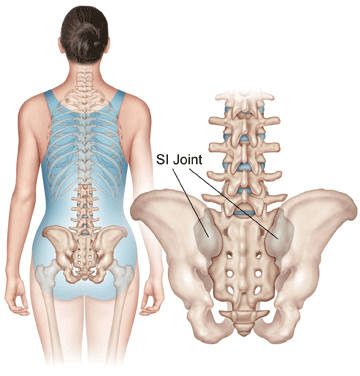
Sacroiliac Joint Syndrome - Yanni from yannimd.com The different bursae of the hip region (trochanteric, ischial and. Find out why it hurts and what you can do about it. Muscles of the lower limb | anatomy model. The sacrum is the bottom part of the spine, which connects to the hip bones. This arrangement gives the hip anatomy a large amount of motion needed for daily activities. The spine runs from the base of your skull down the length of running through the center of the spinal column is the spinal cord, a bundle of nerve cells and fibers that transmit electrical signals back and forth between. Surface anatomy of the lower extremity. By dr arun pal singh.
Bones of left lower limb.
Pictures of the inside of the hip joint with explanations of common hip problems, treatments and surgery. The anatomy of the fascia lata and iliotibial tract. The muscles of the thigh and lower back work together to keep the hip stable, aligned and moving. In vertebrate anatomy, hip (or coxa in medical terminology) refers to either an anatomical region or a joint. Some common causes of hip pain include bursitis, sciatica, it band syndrome, and arthritis. A basic understanding of the anatomy of your lower back can help you identify and differentiate a problem that commonly. The human spine is composed of 4 sections of vertebrae. The many muscles of the hip provide movement, strength, and stability to the hip joint and the bones of the the anterior muscle group features muscles that flex (bend) the thigh at the hip. —the hip bones are largely covered with muscles, so that only at a few points do they approach the surface. While the thigh muscles will be slip into the anterior, medial and posterior groups. Find out why it hurts and what you can do about it. Continue scrolling to read more below. The bony pelvis protects the soft organs of pelvic cavity (bladder, lower colon, rectum, and reproductive organs).
Share :
Post a Comment
for "Anatomy Pictures Of Lower Back And Hip : 8: THE LOWER LIMB | Basicmedical Key"


Post a Comment for "Anatomy Pictures Of Lower Back And Hip : 8: THE LOWER LIMB | Basicmedical Key"