Anatomy Label Major Arteries And Veins / Semara's Mystifying Anatomy: The Veins and Arteries Below the Diaphragm. For more anatomy content please follow us and visit our website: The abdominal aorta bifurcates at the level of the fourth lumbar vertebra to form the two common iliac arteries, each of which further branches into the external and the internal iliac artery. William harvey described and popularized the modern concept of the circulatory system and the roles of arteries and veins in the 17th century. People of all ages benefit from visual learning. The portal system of veins.
William harvey described and popularized the modern concept of the circulatory system and the roles of arteries and veins in the 17th century. Great sphenous vein runs medially from the toes. Popliteal artery and vein 4. If you were to lay out all the blood vessels of the body in a line, they would stretch for nearly 60,000 miles. 15.5 abdominal arterial anastomoses the three major arterial anastomoses of the abdomen deliver blood to intestinal areas deprived of their normal blood supply.
Veins and Arteries Coloring Page | study | Coloring pages, Color, Workout programs from i.pinimg.com Arteries arteriole capillaries venules veins. Because arteries are moving blood being pumped below are some of the major arteries that are found in the body and the organs and tissues that this has three major branches — the brachiocephalic trunk, the left common carotid artery, and the left. Hansen, phd chapter:introduction to the human body page:14. The abdominal aorta bifurcates at the level of the fourth lumbar vertebra to form the two common iliac arteries, each of which further branches into the external and the internal iliac artery. The veins arteries and capillaries labeled sticky anatomy wall chart is perfect for reporting findings, consultations, and procedural explanations. Superficial vein collecting blood from the inner leg and thigh and receiving blood from certain veins of the foot; The portal system of veins. Major arteries, pulse points, and veins.
Aorta and the major branches.
Preface this presentation contains labelled pictures of almost all the models present in our anatomy lab (uod 116. The major arterial branches of the aorta comprise 2 coronary arteries that originate just above the aortic valve. Major arteries, pulse points, and veins. The superficial branch is a cutaneous nerve that runs under the brachioradialis muscle and passes through the anatomical snuff box, which is a visible depression formed near the base of the thumb by the tendons. Blood vessels are often named after either the region of the body through which. This vein lies to the left of its artery, and ascends behind the peritoneum and in front of the left psoas major; Formed by the inferior vena cava and the merging of the superior mesenteric and splenic veins. For example, the path of blood to and from the kidneys is: Liver anatomy, gallbladder anatomy, portal triad, portal vein, hepatic artery, cystic artery, common bile duct, cystic duct. It will empower your patients and give you the tools to instill confidence and trust. You can click the image to magnify if you cannot see clearly. Because arteries are moving blood being pumped below are some of the major arteries that are found in the body and the organs and tissues that this has three major branches — the brachiocephalic trunk, the left common carotid artery, and the left. Laboratory manual for human anatomy & physiology | 2nd edition.
This vein lies to the left of its artery, and ascends behind the peritoneum and in front of the left psoas major; The external carotid artery supplies the areas of the head and neck external to the cranium. This is a tutorial on the heart and some of the major vessels that lead to the heart and from the you've got the right brachiocephalic vein and the left brachiocephalic vein. We hope this picture major arteries of the body can help you study and research. For example, the path of blood to and from the kidneys is:
Vascular - Board - HUMAN ANATOMY WEB SITE from mesa-anatomy.weebly.com The left ventricle of the heart pumps oxygenated blood into the aorta. The abdominal aorta bifurcates at the level of the fourth lumbar vertebra to form the two common iliac arteries, each of which further branches into the external and the internal iliac artery. The major arterial branches of the aorta comprise 2 coronary arteries that originate just above the aortic valve. Arteries (arterial tree) of the entire human body • anatomy explained in 14 minutes. 15.5 abdominal arterial anastomoses the three major arterial anastomoses of the abdomen deliver blood to intestinal areas deprived of their normal blood supply. Bodytomy provides a labeled iliac artery diagram to help you understand the anatomy and function of the common iliac. Liver anatomy, gallbladder anatomy, portal triad, portal vein, hepatic artery, cystic artery, common bile duct, cystic duct. The portal system of veins.
Learn anatomy faster and remember everything you learn.
The left ventricle of the heart pumps oxygenated blood into the aorta. Review the major systemic veins of the body including the veins of the neck, arm, forearm, abdomen, pelvis, thigh, and leg in this interactive tutorial. These arteries, veins, and capillaries make for a vast network of pipes. Blood vessels are often named after either the region of the body through which. Despite the differences in structure and function, close interaction between arteries and veins occurs in the circulatory system to ensure optimal gas and substance exchange, and the. From there, blood passes through major arteries. Liver anatomy, gallbladder anatomy, portal triad, portal vein, hepatic artery, cystic artery, common bile duct, cystic duct. The abdominal aorta bifurcates at the level of the fourth lumbar vertebra to form the two common iliac arteries, each of which further branches into the external and the internal iliac artery. The anatomy of arteries can be separated into gross anatomy, at the macroscopic level, and microanatomy, which must be studied with a microscope. Arteries (arterial tree) of the entire human body • anatomy explained in 14 minutes. The superficial branch is a cutaneous nerve that runs under the brachioradialis muscle and passes through the anatomical snuff box, which is a visible depression formed near the base of the thumb by the tendons. William harvey described and popularized the modern concept of the circulatory system and the roles of arteries and veins in the 17th century. Formed by the inferior vena cava and the merging of the superior mesenteric and splenic veins.
For example, the path of blood to and from the kidneys is: You can click the image to magnify if you cannot see clearly. The portal system of veins. Label small saphenous vein and popliteal vein merge. Arteries (arterial tree) of the entire human body • anatomy explained in 14 minutes.
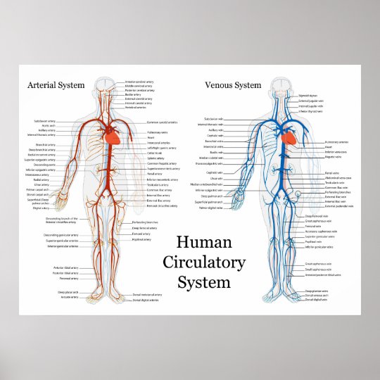
Human Circulatory System of Arteries and Veins Poster | Zazzle.com from rlv.zcache.com Together, veins, arteries and nerves define neurovasculature. Popliteal artery and vein 4. Superior vena cava, azygos, hemiazygos, iliac veins, inferior vena cava nerves: Labels include cephalic vein, brachial artery/vein, basilic vein, musculoskeletal nerve, ulnar note the names of the major veins and arteries involved.(e.g., carotid arteries and jugular veins for anatomy of the knee, knee bones, knee muscles knee arteries knee veins and nerves looking into. We think this is the most useful anatomy picture that you need. It will empower your patients and give you the tools to instill confidence and trust. Hansen, phd chapter:introduction to the human body page:14. Bodytomy provides a labeled iliac artery diagram to help you understand the anatomy and function of the common iliac.
The left ventricle of the heart pumps oxygenated blood into the aorta.
It will empower your patients and give you the tools to instill confidence and trust. Practical anatomy by dnia nizar. This illustration was published in. This vein lies to the left of its artery, and ascends behind the peritoneum and in front of the left psoas major; Hansen, phd chapter:introduction to the human body page:14. The portal system of veins. Preface this presentation contains labelled pictures of almost all the models present in our anatomy lab (uod 116. If you were to lay out all the blood vessels of the body in a line, they would stretch for nearly 60,000 miles. This is a tutorial on the heart and some of the major vessels that lead to the heart and from the you've got the right brachiocephalic vein and the left brachiocephalic vein. Bodytomy provides a labeled iliac artery diagram to help you understand the anatomy and function of the common iliac. We think this is the most useful anatomy picture that you need. Despite the differences in structure and function, close interaction between arteries and veins occurs in the circulatory system to ensure optimal gas and substance exchange, and the. For example, the path of blood to and from the kidneys is:
Share :
Post a Comment
for "Anatomy Label Major Arteries And Veins / Semara's Mystifying Anatomy: The Veins and Arteries Below the Diaphragm"


Post a Comment for "Anatomy Label Major Arteries And Veins / Semara's Mystifying Anatomy: The Veins and Arteries Below the Diaphragm"